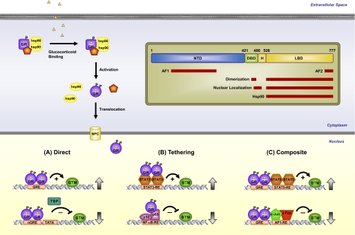
FIGURE 1.
GR signaling pathway. Upon binding glucocorticoids, cytoplasmic GR undergoes a change in conformation (activation), becomes hyperphosphorylated (P), dissociates from accessory proteins, and translocates into the nucleus, where it regulates gene expression. GR enhances or represses transcription of target genes by direct GRE binding (A), by tethering itself to other transcription factors apart from DNA binding (B), or in a composite manner by both direct GRE binding and interactions with transcription factors bound to neighboring sites (C). Inset, GR is composed of an NTD, a DBD, a hinge region (H), and an LBD. Regions involved in transcriptional activation (AF1 and AF2), dimerization, nuclear localization, and chaperone hsp90 binding are indicated. Position numbers are for the human GR. NPC, nuclear pore complex; BTM, basal transcription machinery; TBP, TATA-binding protein; nGRE, negative GRE; RE, response element.

