FIGURE 5.
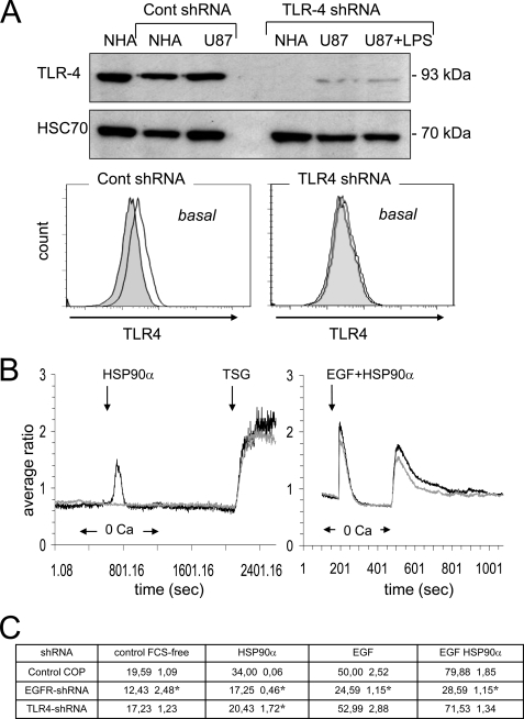
shRNA-mediated depletion of TLR4 inhibits HSP90α-induced Ca2+ signaling in U87 cells. A, protein lysates from both normal human astrocytes (NHA) and glioblastoma U87 cells were probed with anti-TLR4 (HSC70 as loading control). TLR4 expression in normal human astrocytes and U87 cells was shown, and cell stimulation with 1 μg/ml LPS for 60 min was performed. Flow cytometry analysis of TLR4 expression in transfected U87 cells (white histogram) when compared with IgG control (gray filled histogram; n = 3) as performed. B, effects of HSP90α, LPS (1 μg/ml), TSG (2 μm), and HSP90α plus EGF on [Ca2+]i in TLR4 shRNA (grey curves) and control shRNA (black curves) transfected U87 cells (average of 6 cells; n = 3). C, consequences of shRNA-mediated depletion of TLR4 and EGFR (ErbB1) onto the cell invasion. The quantitative effect of HSP90α (6 μg/ml) and/or EGF (10 ng/ml) on the closure of wound after a 12-h treatment is depicted in the table (mean ± S.D., n = 2; *, p values<0.05 versus control). The following cells were used as control in this specific experiment; cultures of U87 cells and normal human astrocytes (Lonza Walkersville, Inc.) were grown to confluency in DMEM plus 10% fetal bovine serum (Lonza). For lentiviral transduction particles and transfections, human TLR4 shRNA, EGFR (ErbB) shRNA and control lentiviral particles were used according to the manufacturer's recommendations (Santa Cruz Biotechnology).

