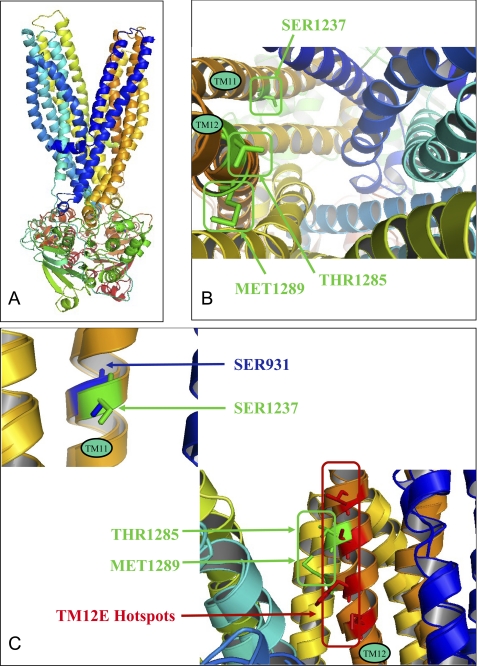
FIGURE 10.
SUR1 structural model and three-dimensional alignment of outward-facing models of P-gp and SUR1. A, outward-facing conformation model of SUR1. B, top view of three-dimensional transmembrane location of residues involved in the binding of KATP channel openers (Thr1285 and Met1289) and blockers (Ser1237). C, structural alignment of TM11 (P-gp)/TM16 (SUR1) and TM12 (P-gp)/TM17 (SUR1). SUR1 residues involved in opener and blocker binding are represented and labeled in green, P-gp TM12E hot spots are in red, and P-gp TM11 residue is in blue.

