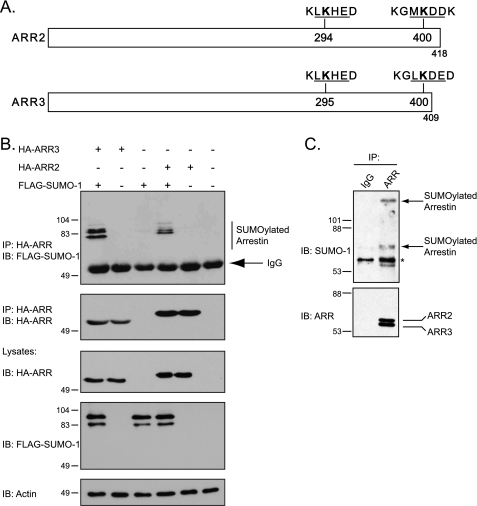
FIGURE 1.
Arrestins are modified by SUMO. A, schematic representation of arrestin-2 and arrestin-3 depicting the putative SUMO consensus sites (underlined) and the surrounding amino acid residues. The SUMO acceptor lysine residues are in bold type. B, SUMOylation analysis of nonvisual arrestins. HEK293 cells were transiently transfected with HA-tagged arrestin-2, HA-arrestin-3, empty vector (pcDNA) plus FLAG-tagged SUMO-1, or empty vector (pCMV) for 48 h. Transfections with empty vector are indicated with a minus sign. HA-tagged arrestins were immunoprecipitated (IP) and followed by immunoblotting (IB) to detect the incorporated FLAG-SUMO-1. The top blot was stripped and reprobed with an anti-HA monoclonal antibody to determine the level of tagged arrestin in the IP. Cross-reactivity to IgG is indicated. The lysates were analyzed by IB to detect the expression of the various constructs. Representative blots from one of three independent experiments are shown. The positions of the molecular mass markers in kDa are shown. C, SUMOylation of endogenous arrestins. Endogenous arrestins were IP from HEK293 cells, followed by IB for endogenous SUMO-1. SUMOylated arrestin is indicated with arrows. The asterisk represents possible cross-reactive bands present in samples incubated with arrestin and IgG antibodies. Representative blots from one of three independent experiments are shown. The positions of the molecular mass markers in kDa are shown.

