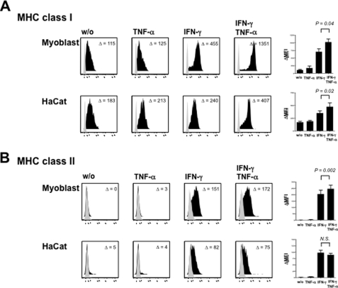
FIGURE 4.
TNF-α increases MHC class I and MHC class II surface expression in IFN-γ-treated primary muscle cells. A, myoblasts and HaCat cells constitutively express HLA class I molecules on their cell surface. MHC class I levels are up-regulated by both TNF-α (50 ng/ml) and IFN-γ (100 ng/ml) and both cytokines act synergistically on MHC class I expression in both cell types. B, IFN-γ induces HLA-DR expression in human myoblasts and HaCat cells, which both lack constitutive MHC class II expression. TNF-α alone does not induce HLA-DR expression on both cell types. In contrast, addition of TNF-α to IFN-γ enhances MHC class II surface expression on myocytes but not HaCat cells. Diagrams display means ± S.E. of MHC expression levels from at least four independent experiments on myoblasts and HaCat cells, respectively. Isotype controls are highlighted in gray. Numbers in each individual histogram represent mean fluorescence intensity compared with isotype control (ΔMFI). MHC expression levels were analyzed using the paired t test.

