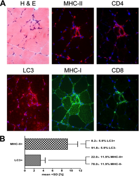
FIGURE 7.
Muscle fibers from patients with sIBM show colocalization of autophagosomes and MHC class II molecules. A, serial and double immunofluorescent staining of a representative skeletal muscle from a patient with sIBM with H&E histochemistry and immunolabeling for LC3, MHC class I and II, CD4, and CD8. Photomicroscopy using a ×40 objective. B, quantitative double-labeling analysis of four patients with sIBM stained for MHC class II and LC3. Values indicate the mean ± S.D. of a total of 1864 skeletal muscle fibers.

