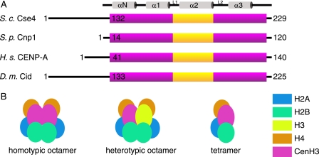
FIGURE 1.
Centromeric nucleosome structure. A, schematic diagram of four CenH3 proteins from S. cerevisiae (S.c.), Schizosaccharomyces pombe (S.p.), Homo sapiens (H.s.), and Drosophila melanogaster (D.m.). The purple bar shows the extent of the histone fold domain, and the yellow box shows the position of the CENP-A targeting domain motif. The secondary structure elements of the histone fold are indicated at the top of the figure. B, possible stoichiometries of centromeric nucleosomes showing the two possible octameric complexes and a tetramer.

