
Figure 1. Polι binary and ternary complexes.
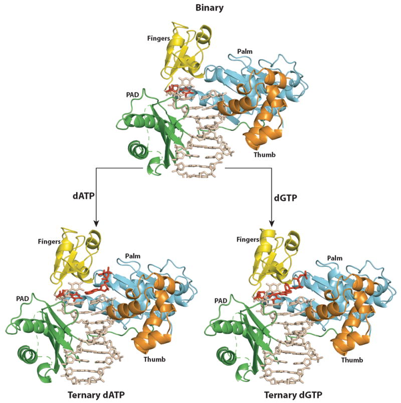
Overall structures of PolιU (top), PolιU.dATP (bottom left) and PolιU.dGTP (bottom right). Palm, fingers, thumb, and PAD domains are shown in cyan, yellow, orange, and green respectively. DNA is shown in tan; the incoming nucleotide and the template U in anti conformation are shown in red. The syn conformation of templating U in the binary structure is shown in gray and has been omitted from the ternary structures for clarity.