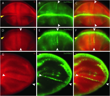
Figure 4.—
N protein levels are elevated at the AP organizer. (A–C) Late third instar wing disc. (D–F) Pupal wing disc. (G–I) Early pupal wing. (A, D, and G) Expression of N protein (red) is high at both the AP (white arrows) and the DV (yellow arrows) border regions. (B, E, and H) Expression of Su(H)lacZ (green) indicates activation of N signaling at both DV and AP borders. (C, F, and I) Merged images show that Su(H)lacZ expression at the AP border is in cells with high levels of N protein.

