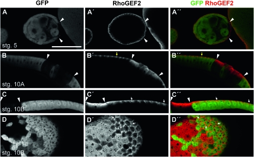
Figure 5.—
RhoGEF2 is basally localized in the follicular epithelium and its localization is regulated by Pak. (A–D) Anti-GFP. (A′–D′) Anti-RhoGEF2. (A″–D″) Merge. FCC are distinguished by a lack of GFP staining. Arrowheads mark some clone boundaries. (A–A″) pak14 FCC in a stage 5 egg chamber showing that the basal localization of RhoGEF2 is slightly reduced. (B–B″) pak14 FCC in columnar cells of a stage 10A egg chamber showing that the localization of RhoGEF2 is no longer restricted to the basal end of follicle cells in the absence of Pak. Yellow arrows mark basal punctate localization of RhoGEF2 in a wild-type cell. (C–C″) pak14 FCC in a stage 10B egg chamber showing ectopic RhoGEF2 distribution throughout cells. (D–D″) pak14 FCC imaged at the basal surface of a stage 10B egg chamber showing that RhoGEF2 accumulation at the points of basal membrane separation is lost in the absence of Pak. White arrows mark obvious sites of basal membrane separation in wild-type tissue. Bar: 50 μm.

