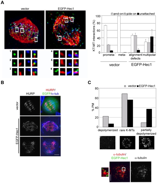
Figure 5. Erroneous kinetochore-microtubule attachments in EGFP-Hec1 cells stabilize K-fibers.
(A) KT-MT attachments in a late prometaphase (vector) and an EGFP-Hec1 expressing disorganized bipolar prometaphase (EGFP-Hec1) exposed to MG132 for 2 h and stained for CREST (blue) and α-tubulin (red) after calcium buffer treatment. Kinetochores (green) are visualized by anti-Hec1 antibody (vector) or EGFP-Hec1 (EGFP-Hec1). Maximum projections from deconvolved z-stacks are shown. Insets show a 3-fold enlargement of 2-3 slices from the boxed regions. In the vector prometaphase two end-on attachments (1,2) and one side-on attachment close to the MT end (3) are shown. In the EGFP-Hec1 cell two side-on attachments similar to the one observed in the vector cell (4,5) and one side-on attachment along a continuing MT (6) are reported. Bar 5 µm. The graph reports a quantitative analysis of the different types of attachments on 175–284 kinetochores in 2–3 cells for each mitotic stage. Alignment defects include disorganized bipolar prometaphases. (B) HURP (red) and α-tubulin (blue) staining in vector or EGFP-Hec1 cells. HURP localization extended towards the MT minus-ends in the EGFP-Hec1 cell. (C) Quantitative analysis of MT depolymerization in NOC-treated vector and EGFP-Hec1 prometaphases (PM) (n = 200). Representative examples of the different classes of MT depolymerization are presented below the graph (α-tubulin staining). Localization of residual MTs (α-tubulin, red) with respect to KTs (EGFP-Hec1, green) in a EGFP-Hec1 expressing prometaphase after NOC treatment. Inset shows a 3-fold enlargement of the boxed region in the merge.

