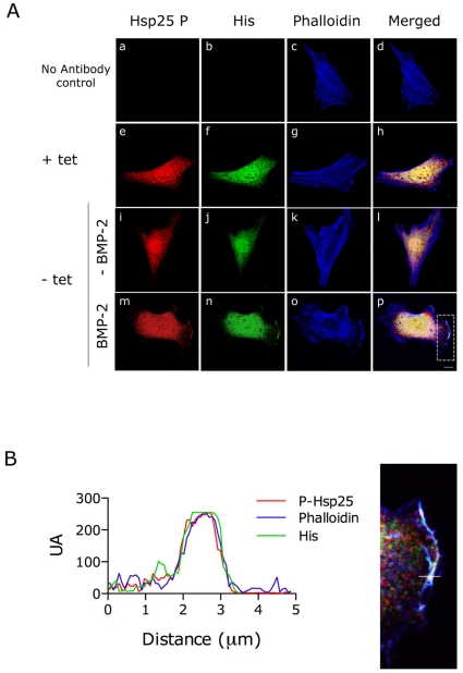
Figure 8. Phosphorylated Hsp25 and BMPRII colocalize in membrane protusions upon BMP-2 stimulation.
(A) Immunostaining of phospho-Hsp25 (red) and His (green) of C2C12 cells overexpressing His-tagged BMPRII under a tetracycline (Tet-off) regulated promoter. Cells were serum-starved for 16 h, pre-treated with cytochalasin D and allowed to recover for 1 h in the absence (i-l) or presence (m-p) of BMP-2. Actin cytoskeleton was stained with Alexa Fluor 633 phalloidin (blue) and merged images are shown. (a-d), Controls where primary antibodies have been omitted from the incubation. (e-h), Immunostaining of the His-tagged BMPRII overexpressing clone incubated in the presence of 30 ng/ml of tetracycline for 24 h showing the diffuse background nuclear staining by anti-His antibody. Scale bar, 40 µm. (B) Spatial quantification was performed along a path across the plasma membrane, indicated by a white line in the detail amplification of the merged image above. Red, green and blue fluorescence was quantified separately and plotted as a function of distance along the path.

