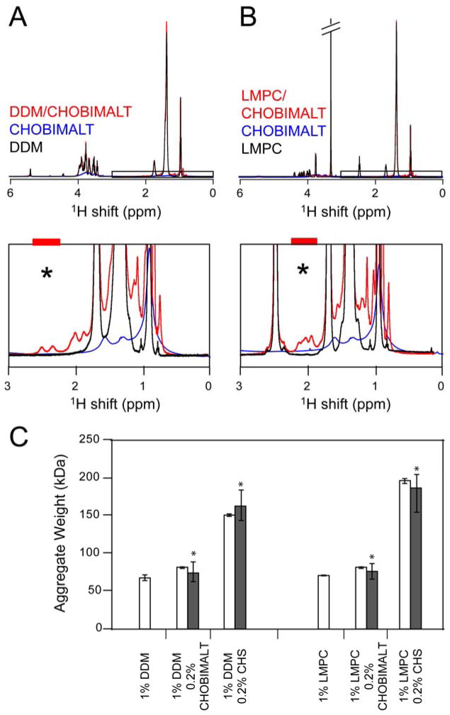
Figure 6.
NMR assessment of the formation of mixed micelles involving detergents and cholesterol analogs. A. Overlay of 1H spectra for 1% (20 mM) DDM, 0.2% (1.9 mM) CHOBIMALT, and 1%/0.2% DDM/CHOBIMALT (ca. 10:1 mol:mol) mixed micelles. B. Overlay of 1H spectra for 1% (21 mM) LMPC, 0.2% (1.9 mM) CHOBIMALT, and 1%/0.2% LMPC/CHOBIMALT (ca. 10:1 mol:mol) mixed micelles. In both mixed micelle systems, incorporation of the cholesterol analog into the smaller detergent micelle gives rise to the observance of resonances (1.8–2.8 ppm) not clearly seen in the spectra of the pure cholesterol analogs. Diffusion measurements were made on isolated resonances defined by the colored bars above the black asterisk, with the CHOBIMALT- or CHS-specific diffusion coefficients being compared to the results obtained by integrating over the aliphatic protons, which are dominated by signal from the detergent. C. Influence of CHOBIMALT and CHS on the size of detergent micelles. Aggregate masses were determined from translational diffusion coefficients measured as indicated in B, either on the aliphatic protons (white bars) or the isolated sterol resonances (black bars).

