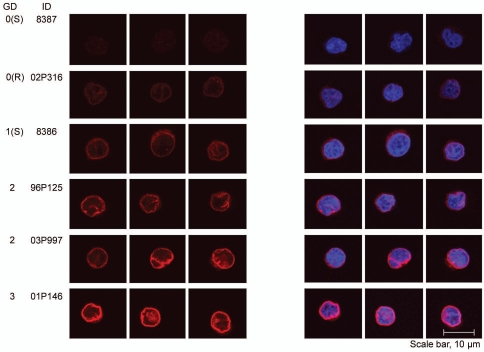
Figure 5.
Dose dependent effect on the staining intensity of the nuclear membrane in lymphoblastoid cells in immunofluorescence microscopy using an anti-LBR antibody. A representative set of LCLs is shown. To evaluate and compare LBR staining intensities, confocal images were collected with identical laser intensities and scanning parameters.

