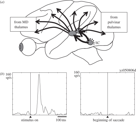
Figure 5.
Contribution of the SC—pulvinar—cortical pathway to the visual suppression with saccades. (a) Symmetry of projections from MD to frontal cortex and from pulvinar to parietal and occipital cortex. (b) Increased activity of a pulvinar neuron in the path from SC to MT to a visual stimulus (left), and decrease in activity of that neuron following a saccade (right). Adapted from Berman & Wurtz [53].

