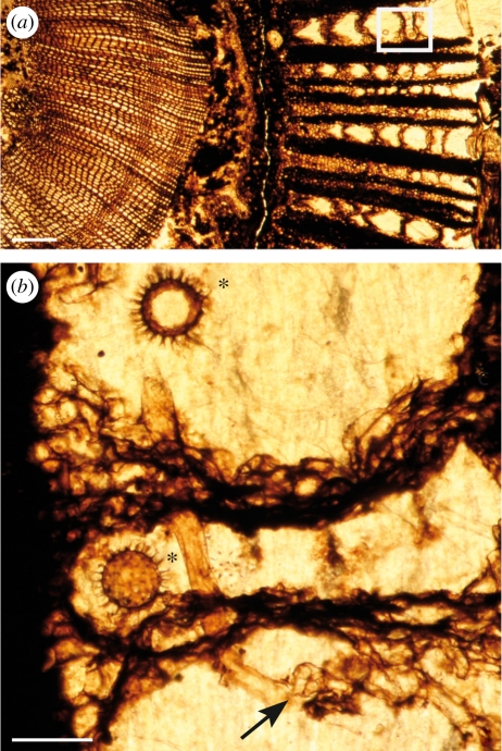
Figure 1.
(a) Overview of a transverse section of Lyginopteris oldhamia stem showing colonization by the Oomycetes in the cortical tissues (frame); the zone in the frame corresponds to (b); scale bar, 400 µm. (b) Hypha (arrow) and oogonia (asterisk) of Combresomyces williamsonii (Holotype) within the parenchyma that separates the fibres of the dictyoxylon outer cortex of the stem. Note the occurrence of a knot of hyphae (arrow); scale bar, 130 µm. All images from slide specimen NHMUK PB.WC.1144.E.

