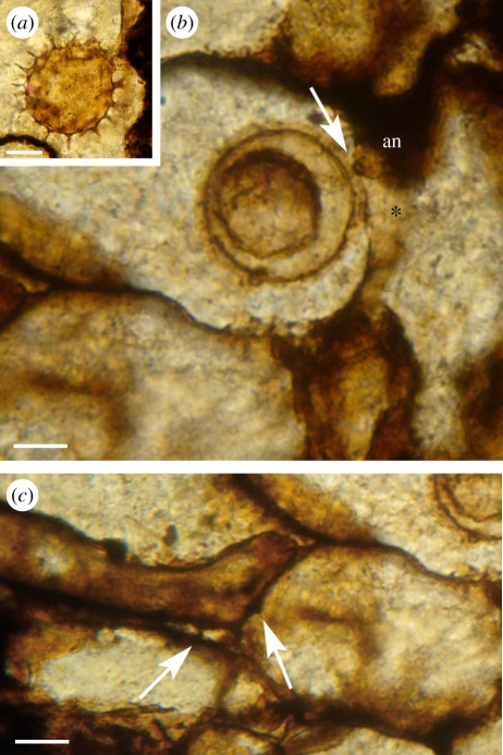
Figure 3.
Combresomyces sp. (a) Oogonium in the parenchymatous intercalation of the stem cortex of Lyginopteris oldhamia; a smaller sphere is discernible on the inside of the oogonium; scale bar, 25 µm. Slide specimen MANCH R.1077. (b,c) Colonization in Lyginopteris stem (in longitudinal section). (b) Note the fertilization tube in the oogonium/antheridium complex (arrow) and the aplerotic oospore; antheridium (an) and antheridium stalk (asterisk); scale bar, 15 µm. (c) Vegetative hyphae developing possible haustoria (arrows) that penetrate the cell walls of the host but not the cytoplasm; scale bar, 15 µm. Slide specimen MANCH R.1081.g.

