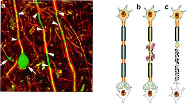
Figure 1. Immune-mediated demyelination and axonal transection.
Axonal ovoids are hallmark of transected axons. Abundant axonal ovoids were detected in MS tissue (a) when stained for myelin protein (red) and axons (green). There are areas of demyelination (arrowheads), mediated by microglia and hematogenous monocytes. One of the axons ends in a large swelling (arrow) or axonal retraction bulb (arrow). (b-c) Schematic of axonal response during and following transection. Demyelination is an immune-mediated or immune cell assisted process leading to axonal transaction. When transected, the distal end of the axon rapidly degenerates while the proximal end connected to the neuronal cell body survives and transported organelles accumulate at the transection site and form an ovoid (arrows). (Reproduced from(Trapp and Nave, 2008)

