
Fig. 1.
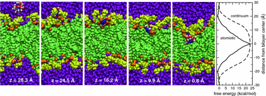
The first five panels show snapshots of a guanidinium ion passing from bulk water to the middle of a phospholipid bilayer (Gdm blue; water in solvation shell red O, white H; lipid carbonyl orange; phosphocholines yellow; water purple). The ion center-of-mass distance from the center of the bilayer along the bilayer normal is indicated at the bottom of each panel. The rightmost panel shows the free energy profile (PMF) for the Gdm ion in a lipid bilayer computed by atomistic MD simulations (solid line), and the electrostatic free energy (dashed line) in a continuum model of the membrane as a rigid slab (dielectric constant εM = 1) with sharp interfaces at z = ±20 Å with semi-infinite regions of water (εW = 89), including both the ion self-energy contribution, given by the Born solvation formula, and that due to the polarization of the slab by the charge. The PMF was mapped from z = 0 (bilayer center) to z = 30 Å, where it was set to 0. Given the system symmetry, at equilibrium the PMF should be symmetric with respect to the center of the bilayer. We therefore reflected the PMF about z = 0 for clarity of presentation. In the continuum calculation we adjusted the radius of the charge to match the PMF at z = 0. The resulting value of 5.7 Å compares reasonably well with the average location of the first coordination shell (~5 Å from the Gdm carbon atom) (Color figure online)