
Fig. 5.
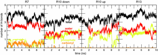
Time dependence of the number of H bonds between Arg side chains and water (red), and lipid phosphate (yellow) and carbonyl (yellow) groups in helical TM configurations of the GGPGL6RL13GPGG (R7), GGPGL9RL10GPGG (R10), and GGPGL12RL7GPGG (R13) peptides in a POPC bilayer. Two initial conditions (“up” and “down”) were used for the R10 peptide, in which the Arg residue is in the middle of the sequence of the TM segment. The Arg residue participates in 3–4 H bonds at all times in each case (Color figure online)