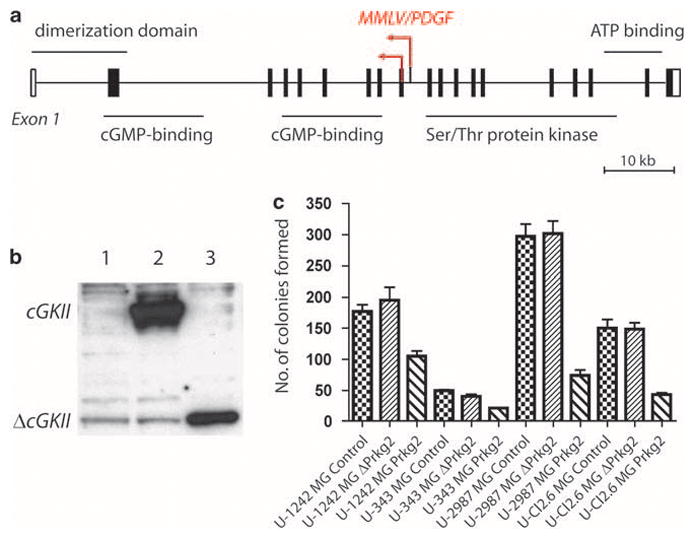
Figure 1.

(a) Schematic picture of the two proviral integrations of Moloney murine leukemia virus (MMLV)/platelet-derived growth factor (PDGF) in Prkg2 on mouse chromosome 5. Major structural domains of the protein are presented. Data were obtained from National Center for Biotechnology Information, Mouse 37 assembly and ENSEMBL Release 51, 2008. (b) Transiently transfected Cos-1 cells (after 72 h) overexpressing an empty pcDNA vector (lane 1), full-length cGKII (lane 2, ~85 kDa) or truncated ΔcGKII (lane 3, ~35 kDa). (c) Colony-forming assay showing a number of colonies formed in four different glioma cell lines (U-1242MG, U-343MG, U-2987MG and U-Cl2:6MG) transfected with Prkg2, ΔPrkg2 or empty pcDNA (Control). Colonies were counted after 3 weeks of growth in medium containing 0.8 mg/ml of neomycin. The cells were plated in triplicates and experiments were repeated twice. Results are presented from one representative experiment and significant differences were found for Prkg2 transfections compared with empty vector transfections (P<0.05) with paired t-test for every cell line, but not for ΔPrkg2 and empty vector in U-Cl2:6MG. The U-87MG cell line yielded too few colonies after transfection and was not included.
