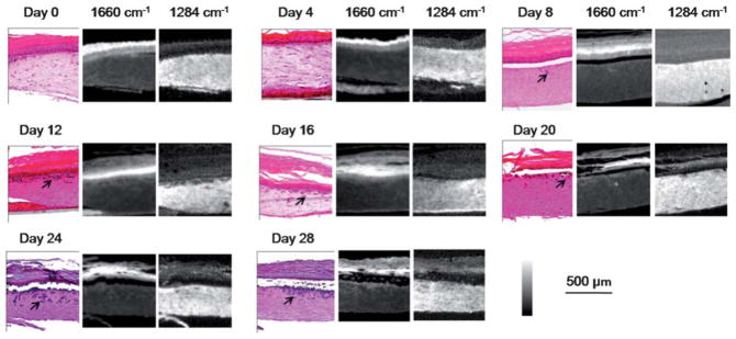
Fig. 2.
Melanoma progression in 28 days. Tumors (arrows) grow larger and invade into the dermis over time, as shown by both H&E-stains followed by histological recognition and IR spectroscopic imaging. IR absorbance images at 1660 cm−1 and 1284 cm−1 highlight epidermal and dermal regions, respectively, indicating different chemical compositions between these two regions and their facile delineation by simple spectral metrics.

