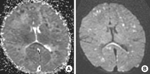
Fig. 3.
Follow-up MRI obtained on 10th day of birth. (A) ADC showed low-signal-intensity lesions in the corresponding areas. (B) DWI showed more decreased signal-intensity of previous lesions and newly developed high-signal-intensity lesions in both cerebral hemispheres, especially in the periventricular white matter and corpus callosum.

