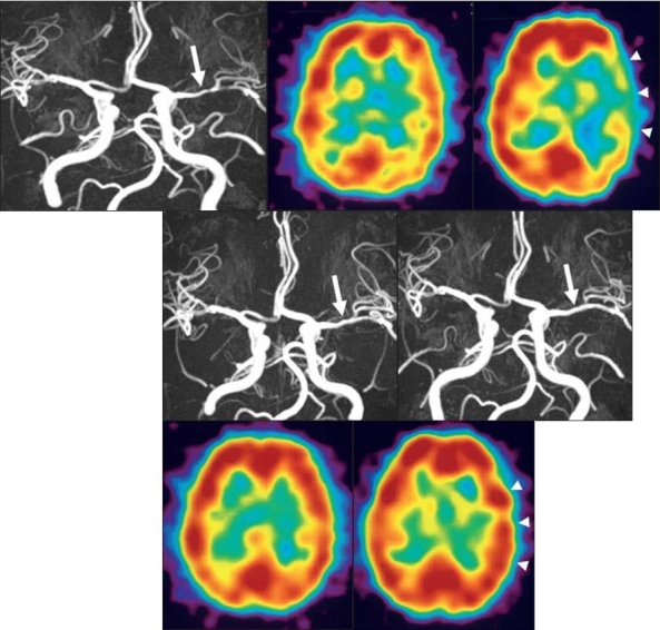
Figure 3.

3A: Pretreatment MRA shows a stenotic lesion (85%) at the left M1 (white arrow)
3B: Pretreatment SPECT shows no laterality in CBF at rest.
3C: Pretreatment SPECT shows hypovasoreactivity in the left temporo-occipital region after acetazolamide challenge (white arrow head).
3D: MRA obtained 6 months after the start of cilostazol treatment shows improvement of the stenotic lesion (30%) at the left M1 (white arrow).
3E: MRA acquired 12 months after the start of cilostazol treatment shows improvement of the stenotic lesion (20%) at the left M1 (white arrow).
3F: SPECT obtained 12 months after the start of cilostazol treatment shows increased CBF in the left cerebral hemisphere.
3G: SPECT obtained 12 months after the start of cilostazol treatment shows CBF improvement in the left cerebral hemisphere after acetazolamide challenge (white arrowhead).
