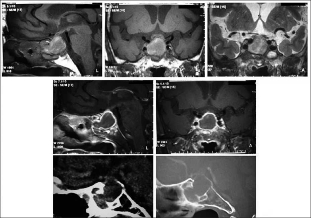
Figure 2.

Magnetic resonance images and CT images after neurological and hormonal symptoms appeared. Upper left and center: T1-weighted images, upper right: T2-weighted image. Middle left and center: T1 weighted images after administration of contrast medium showing enhancement of the outline of sellar lesion. Lower left and center: CT image showing intra- and supra-sellar enhanced mass lesions, defect of the sella floor, and extension of the pituitary lesion toward the shpenoid sinus.
