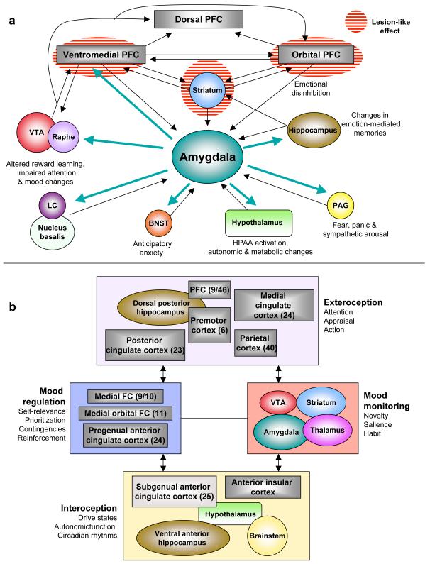
Figure 1. Two heuristic formulations of “depression circuits.”.
a) An amygdala-centric circuit (8) largely inspired by structural brain imaging and postmortem studies. According to this model, the emotional symptoms of depression can be brought about by functional impairment (“lesion-like” effects) of the striatum or prefrontal and orbital prefrontal cortex (and/or their associated white matter tracts), resulting in disinhibition of the amygdala and downstream structures. Alternatively, they can arise from functional hypersensitivity of the amygdala (turquoise arrows) which gives rise to dysregulation of prefrontal cortical structures. b) Another circuit model (138) generated with a greater emphasis on functional imaging results. The main nodes consist of four clusters of brain regions with strong anatomical connections to each other; bidirectional arrows indicate strong inter-connections. This model compartmentalizes depressive endophenotypes into exteroceptive (cognitive), interoceptive (visceral-motor), mood-regulating and mood-monitoring functions, with numbers in parentheses reflecting formal Brodmann area assignments. Both formulations should be seen as offering a simplified heuristic framework for further research into depression’s diagnosis, pathophysiology and treatment. They do not convey the cellular and molecular heterogeneity of each node within the circuit (for example, the VTA is comprised of several types of dopaminergic and GABAergic neurons defined by differences in connectivity and receptor expression). Abbreviations: VTA (ventral tegmental area), LC (locus coeruleus), BNST (bed nucleus of the stria terminalis), PFC (prefrontal cortex), PAG (periaqueductal gray).

