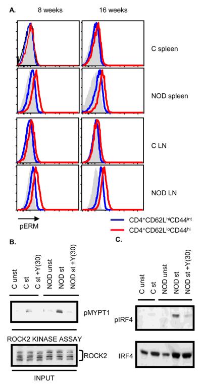
Figure 1.
Enhanced ROCK2 activation and IRF4 phosphorylation in NOD CD4+ T cells. A. Increased levels of phosphorylated Ezrin/Radixin/Moesin (pERM) proteins in NOD CD4+ T cells. The levels of pERM in CD4+CD44intCD62Lhi (blue line) and CD4+CD44hiCD62Llo cells (red line) from either Balb/c or NOD mice were detected by intracellular staining with an anti-phospho-ERM antibody and analyzed by FACS. Solid histograms indicate isotype control. Results are demonstrated as overlaid histograms after gating on the appropriate populations. Data are representative of two independent experiments. B. Purified CD4+ T cells from Balb/c (C) or NOD mice were either left unstimulated or restimulated with αCD3 and αCD28 mAbs for 48 hours in the presence or absence of 30μM of Y27632 (Y(30)). ROCK2 kinase activity was assayed by incubating the immunoprecipitated ROCK2 with purified recombinant MYPT1 (rMYPT1) as ROCK2 substrate. Phosphorylated rMYPT1 (pMYPT1) was detected using an anti-phospho-MYPT1 antibody (upper panel). Lower panel shows the total ROCK2 levels in the input samples. C. CD4+ T cells from Balb/c (C) or NOD mice were stimulated as described in Figure 1B. Nuclear extracts were analyzed by Western blotting using an antibody that recognizes phosphorylated IRF4 (pIRF4) (upper panel). The blot was later reprobed with an antibody against total IRF4 (lower panel). The data are representative of three independent experiments.

