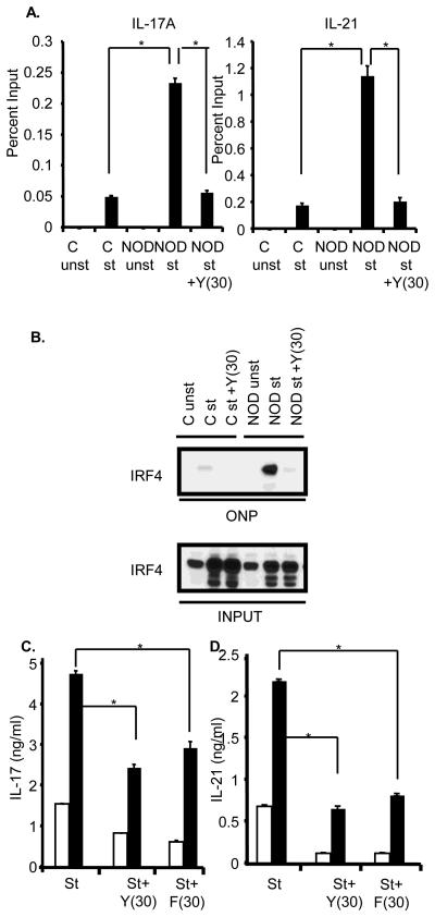
Figure 2.
ROCK inhibition interferes with IRF4 function and diminishes the production of IL-17 and IL-21 by NOD CD4+ T cells. A. CD4+ T cells from either Balb/c (C) or NOD mice were cultured as described in Fig. 1B. Chromatin immunoprecipitation assays were then carried out with either an IRF4 antibody or a control antibody. Quantification of IRF4 binding to the IL-17A and IL-21 promoters was performed using QPCR and is shown as percent input. The data are representative of two independent experiments. The error bars represent Mean ± S.D. *P≤0.0008. B. CD4+ T cells were purified from Balb/c (C) or NOD mice, were cultured as described in Fig. 1B, and then subjected to an oligonucleotide precipitation assay (ONP) with a biotin-labelled oligonucleotide corresponding to the IRF4 binding site within the IL-21 promoter. Precipitates were analyzed by Western blotting with an IRF4 antibody (top panel). The levels of IRF4 in the input samples are shown in the lower panel. Data are representative of three independent experiments. C. Purified CD4+ T cells from Balb/c (white bars) and NOD (black bars) mice were subjected to a primary stimulation and then restimulated with αCD3 and αCD28 for 48 hrs in presence or absence of 30 μM of Y27632 (Y(30)) or 30 μM of Fasudil (F(30)). Supernatants were assayed for IL-17 (left panel) and IL-21 (right panel) production by ELISA. The data are representative of three independent experiments. The error bars represent Mean ± S.D. *P≤0.004.

