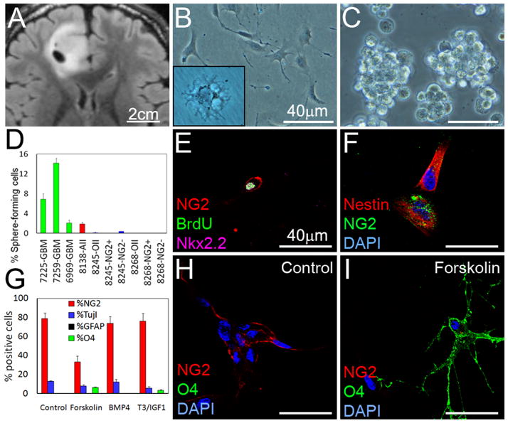
Figure 8. Sphere formation and differentiation potential of human oligodendroglioma cells.
Cells were isolated from a human 1p/19q deleted oligodendroglioma (T2-weighted FLAIR image in A, SF#8245) and cultured as adherent (M41 media in insert, B) or clusters (C). (D) Sphere formation potential in NG2+ and NG2− cells from 2 acutely isolated 1p/19q deleted oligodendrogliomas. (E–F) Incubation of cells from (A) with BrdU 2h before fixation, followed by staining for OPC-related genes NG2 and Nkx2.2, and stem cell marker Nestin. (G–I) To investigate differentiation, we incubated tumor cells that had been passaged once, with forskolin, BMP4, or T3/IGF1 (7d). Quantification (G) with representative images (H–I) demonstrating staining with NG2 and O4 in response to forskolin. Values are expressed as mean ± SEM. See also Figure S7.

