
FIGURE 3.
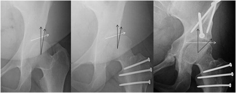
Anteroposterior hip radiographs. Preoperative radiograph on the left demonstrates a method for determining acetabular coverage when measuring center edge angles in hips with aspherical femoral heads associated with Perthes disease. Postoperative radiograph on the right utilizes the same technique when determining the center trochanteric distance, thedistance in mm that the trochanter is above (negative) or below (positive) the center of the femoral head.