FIGURE 4.
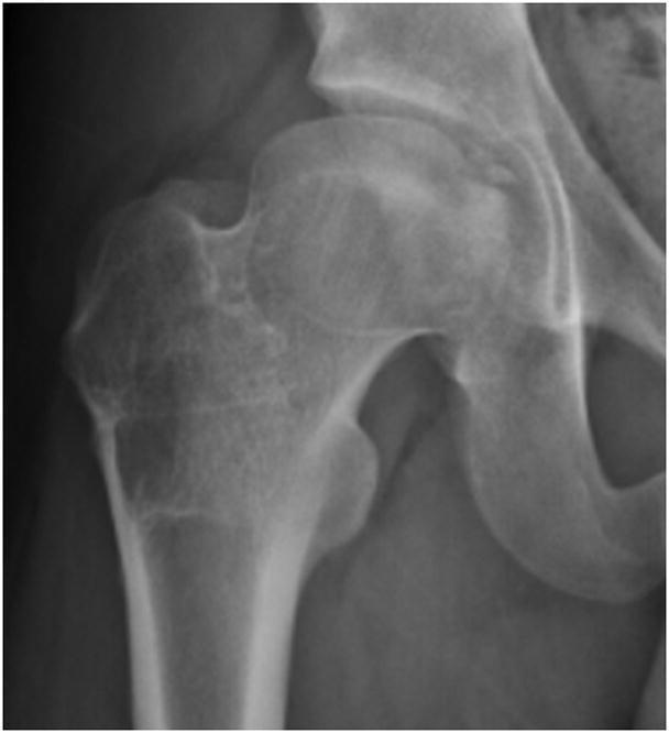
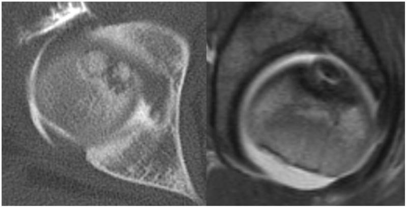
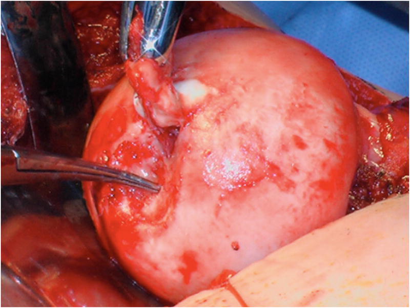
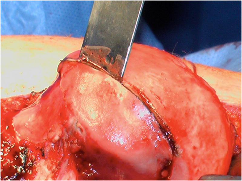
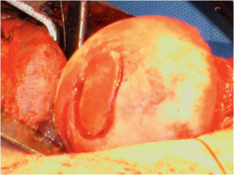
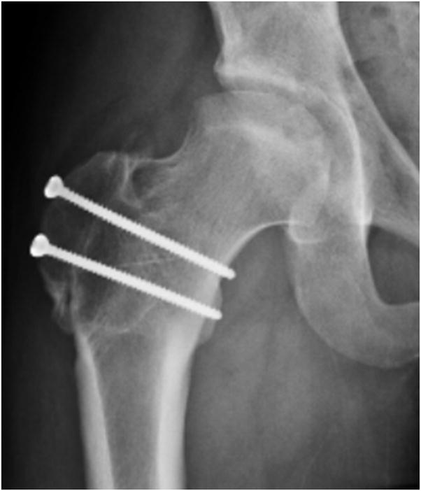
A–F, Preoperative MR arthrographic and radiographic images of a femoral OCD lesion and intraoperative pictures of the subsequent treatment. A, Preoperative anteroposterior radiographic demonstrating reduced CTD, a mushroom shaped femoral headand femoralOCD lesion. B, Preoperative sagittal (left) and axial (right) MR arthrogram images demonstrating a femoral OCD lesion. C, Intraoperative photographs of a femoral head OCD lesion undergoing debridement. D, Intraoperative photographs of anosteochondroplasty of the femoral head-neck juntion being performed which will be used as the osteochondral autograft for the femoral head OCD lesion. E, Intraoperative photographs and postoperative radiographs of an osteochondral autograft of a femoral head OCD lesion. F, Postoperative anteroposterior radiographic demonstrating improved CTD and head-neck offset, and healing off emoral OCD lesion. CTD indicates center trochanteric distance; OCD, osteochondral defect; MR, magnetic resonance.
