
Figure 1. Unliganded Model of the HIV-1 Envelope Trimer.
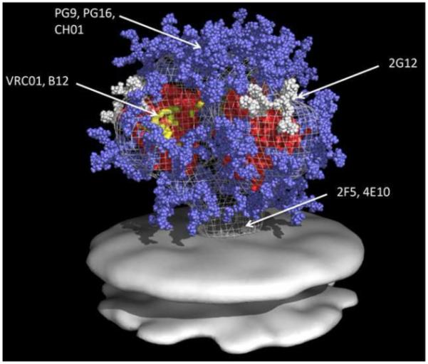
All glycans on gp160 are shown. Glycans are light blue and white, core gp120 is red, 2G12 epitopes are white (arrow), B12 and VRC01 epitopes are yellow (arrow), and the location of the quarternary epitope involving V2 and V3 loops of mAbs PG9 and PG16 is indicated. Image courtesy of Dr. William Schief, University of Washington, Seattle, WA, adapted with permission from (Schief et al., 2009).