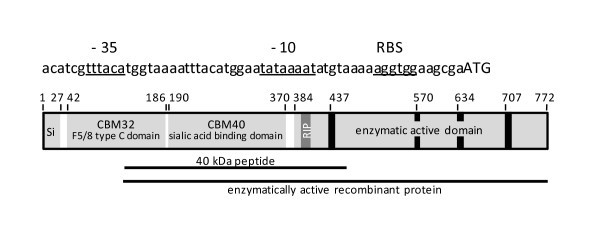
Figure 1.

Schematic representation of the NanA sialidase of C. chauvoei. Upper part: DNA sequence of the promoter region of nanA of strain JF4135 with -35 and -10 boxes and the ribosome binding site (RBS) underlined. The translational start codon (ATG) is indicated by upper case letters. Lower part: main domains of the NanA polypeptide. Numbers refer to amino acid positions and black bars represent the characteristic "Asp-boxes". Si is the signal sequence and RIP indicates the position of the first Arg residue of the catalytic triad with the characteristic Arg-Ile-Pro sequence. The horizontal lines indicate the part of NanA that was expressed as a recombinant 40 kDa polyhistidine-tailed peptide to produce monospecific polyclonal antibodies in rabbits (upper) and the part of NanA expressed as enzymatically active recombinant protein (lower).
