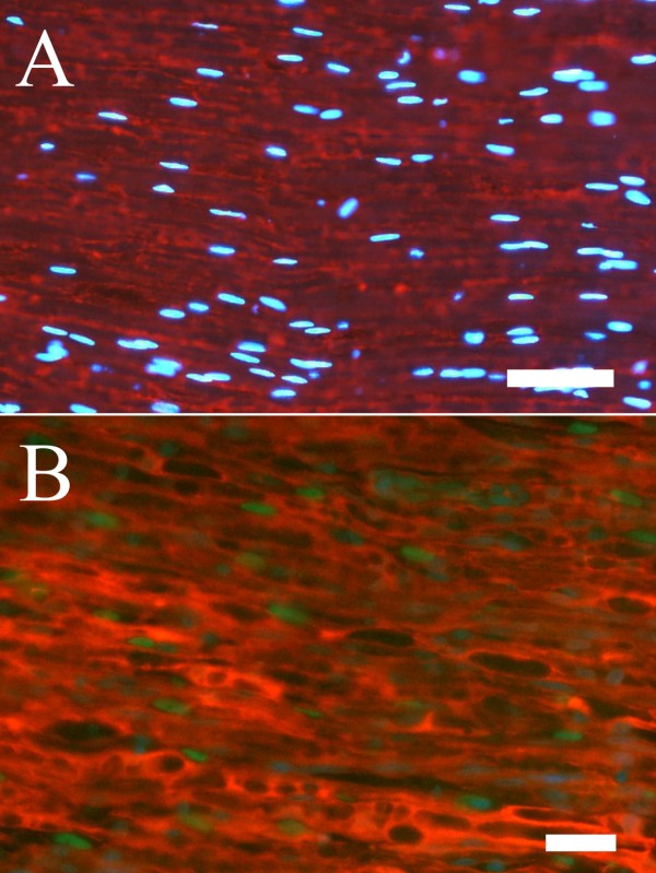
Figure 2.
Double staining with S-100 and cleaved caspase 3 in a control nerve (A) and in a repaired (B) sciatic nerve from the distal nerve segment. The staining indicated that cleaved caspase 3 stained cell nuclei (green) was associated with S-100 staining (red). DAPI-stained cells (blue) were used to localize cell nuclei. Length of bars 50 μm (A) and 25 μm (B).

