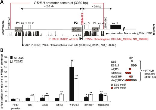
Figure 6.
PTHLH promoter characterization and PTHLH reporter gene assays in chondrocyte cell lines. (A) 5′ RACE PCR of PTHLH-transcripts in ATDC5 and C28/I2 cells. Two TATA boxes (P1, P3), one GC-rich promoter (P2), the UCSC mammalian conservation with approximate genomic kilobases distances and the PTHLH exons 1–5 (ex.) are shown. Identical chondrogenic transcription start site (TSS, bp 28014179) were revealed in ATDC5 and C28/I2 cells downstream of GC-rich promoter (bottom). Two further PTHLH transcripts start at bp 28016183 (see NCBI RefSeq). The red box represents 3080 bp of the PTHLH promoter used to generate the reporter construct. (B) PTHLH-luciferase reporter gene assays with EBS (C-ets-1 core consensus sequence), wt(12) (AP-1 motif), der(8) BP (C-ets-1 and AP-1 motif) or a three time repeats (×3) inserted upstream of the 3080 bp-PTHLH promoter (scheme right). Only the wt(12) × 3 permitted strong stimulation of PTHLH promoter activity and the presence of the C-ets-1 motifs in der(8) BP × 3 prevented activation by endogenous factors (n = 3, **P < 0.01, *P < 0.05).

