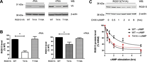
Figure 5.
Crucial role of Thr41 in the maintenance of RGS13 protein expression in vivo. (A and B) pcDNA3.1/V5-His RGS13 (WT or mutants) plasmids were transfected into HEK293T or HeLa cells in the presence or absence of a plasmid encoding the catalytic subunit of PKA. After 24 h, cell lysates were prepared and analyzed by immunoblotting as indicated. Bar graphs in (B) show the mean ± SEM (fold of RGS13 WT) of three independent experiments (*P < 0.005, one-way ANOVA, RGS13 WT vs. T41A). (C) Critical role of Thr41 in cAMP-induced inhibition of RGS13 decay. HEK293T cells were transfected with pEGFP-RGS13-T41A. Twenty-four hours later, cells were stimulated with 8-pCPT-cAMP for the indicated time periods in the presence of CHX followed by collection of cell lysates and immunoblotting by anti-RGS13 and anti-β-actin antibodies (upper panel). Densitometric analysis of RGS13 levels (normalized to β-actin) are shown in from the lower panel (mean ± SEM of four independent experiments; P < 0.01, RGS13 WT + cAMP vs. RGS13(T41A) + cAMP, Student's t-test. The decay curve of RGS13(T41A) is overlaid with curves from Figure 3B for comparison.

