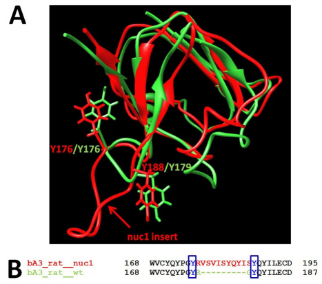
Fig. 11.
Superposition of C-terminal domains of wild-type rat βA3-crystallin and Nuc1 mutant protein. (A) Polypeptide ribbons for wild-type rat βA3-crystallin and the Nuc1 mutant are shown in green and red, respectively. The putative tyrosine-based sorting signal GYxxϕ is located at positions 175–179 on the protein surface. Tyrosines 176 and 179 are shown in green. The tyrosine-based sorting signal GYxxϕ is interrupted by the ten-residue insert located between Y176 and Y188 in the Nuc1 mutant protein (red). (B) The sequence alignment of wild-type rat βA3-crystallin and Nuc1 mutant protein. The positions of the two tyrosine residues from the putative tyrosine-based sorting signal GYxxϕ are shown in blue.

