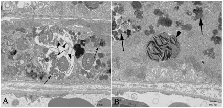
Fig. 4.
At higher magnification, prominent features of RPE cells from 1-year-old Nuc1 animals are shown. (A) An RPE cell showing large aggregates of lipofuscin-like material. A large vacuole contains many degenerated cellular organelles (arrowheads) intermixing with lipofuscin-like aggregates (arrows). (B) Abundant lipofuscin-like aggregates (arrows) and a large phagosome containing undigested outer segment discs (arrowhead) are present in the cytoplasm of an RPE cell. Scale bars: 500 nm.

