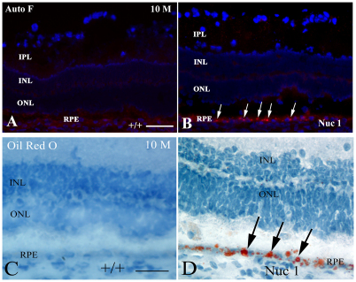
Fig. 5.
Demonstration of abnormal lipid accumulation in the Nuc1 RPE. (A,B) At 10 months of age, minimal autofluorescence is seen in the RPE cells of the wild-type animals (A). However, abundant autofluorescence (arrows) is observed in the RPE of 10-month-old Nuc1 animals, consistent with accumulation of lipofuscin (B). (C,D) Autofluorescence viewed with a red filter (560–620 nm). Oil Red O staining, indicating the presence of neutral lipids, shows abundant intense staining in the RPE cells of the 10-month-old Nuc 1 retinas (D, arrows), but not in the control wild-type animals (C). Scale bars: 50 μm.

