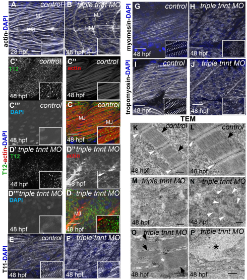
Fig. 1.
Disruption of sarcomere assembly of fast-twitch muscle of embryos simultaneously depleted of tnnt2c, tnnt3a and tnnt3b. (A,B) Phalloidin staining shows forming and mature striated myofibrils in control embryos at 28 hpf (A) but completely disrupted myofibrils in triple tnnt MO-injected embryos at the same stage (B). MJ, myotendinous junctions; HM, horizontal myoseptum. (C,D) At 48 hpf, the titin epitope T12 (C′,D′; green) localises to normal Z lines, and actin (C″,D″; red) appears in striations in control embryos (C), whereas the two proteins are randomly dispersed in the cytoplasm in triple tnnt MO-injected embryos (D). DAPI stains the nuclei (C‴,D‴; blue). Single channels are shown in grey. (E–H) The titin epitope T11 localises to the I-band–A-band boundaries and myomesin to the M lines in control embryos (E,G). They are still seen in discrete structures, although these are scattered around in the cytoplasm, in triple tnnt MO-injected embryos (F,H). (I,J) Tropomyosin localisation to thin filaments observed in the control (I) is lost in the triple tnnt MO-injected embryos (J). Scale bars: 25 μm, insets are 2× greater magnifications of corresponding panels. (A,B) Lateral views of rostral somites of 28 hpf embryos. (C–J) Lateral views of a single myotome. Confocal images were obtained from planes deep in the trunk musculature. (K–P) TEM images of fast-twitch muscles in 48 hpf control (K,L) and in triple tnnt MO-injected embryos (M–P). White arrowheads indicate Z lines and black arrows indicate M lines. In M, clusters of thick filaments can be observed. Accumulations of Z line proteins can be seen in N; in O, thin filament-free sarcomeres can be observed. The white asterisk marks the area where the Z line should have been. Accumulations of actin can be observed in P (black asterisk). Scale bars: 500 nm.

