Fig. 2.
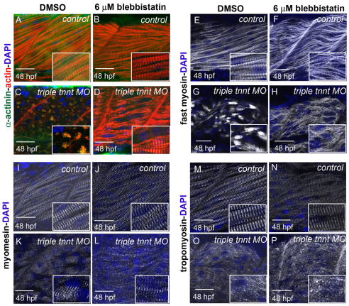
Recovery of striated myofibrils in triple tnnt-depleted embryos upon inhibition of myosin activity. (A–L) Phalloidin (red in A–D), α-actinin (green in A–D), fast myosin (white in E–H) and myomesin (white in I–L) staining of embryos incubated with DMSO reveals normal myofibrils in a control embryo (A,E,I) and the expected disruption in distribution of actin, Z lines, thick filaments and M lines in triple tnnt MO-injected embryos (C,G,K). When embryos are treated with blebbistatin, less compact myofibrils are observed in controls (B,F,J) and striated myofibrils can be seen in triple tnnt MO-injected embryos (D,H,L). (M–P) Tropomyosin does not recover its localisation to thin filaments in tnnt MO-injected embryos treated with blebbistatin (P). DAPI (blue) stains the nuclei. Scale bars: 25 μm, insets are 2× magnifications of corresponding panels.

