Fig. 5.
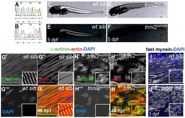
Progressive loss of fast-twitch fibre myofibrillar integrity in tnni2sa0058 mutant embryos. (A,B) Sequence traces from a wild-type fish (A) and a tnni2a.4sa0058 carrier fish (B) show the nucleotide change (asterisk) that converts C to T, generating a stop codon. (C–F) In a 5-day-old wild-type sibling observed under polarised light (C,E, at two different light intensities) trunk muscles are birefringent, whereas in a mutant embryo they are not (D,F). (G,H) Lateral views of single myotomes. Confocal images were obtained from planes deep in the trunk musculature. At 48 hpf, tnni2a.4 mutant embryos show myofibrils that are losing their integrity, as highlighted by phalloidin staining for thin filaments (G″,H″; red) and α-actinin staining for Z lines (G′,H′; green). DAPI stains the nuclei (G‴,H‴; blue). Single channels are shown in grey; overlap of signals shows in yellow in the merged panels (G,H). Scale bars: 10 μm. (I,J) At 3 dpf, fast myosin (white) is completely delocalised in tnni2a.4 mutants (J) whereas wild-type siblings show normal fast muscle (I). Nuclei are stained in blue. Scale bars: 20 μm. Insets are 2× magnifications of corresponding panels.

