Fig. 8.
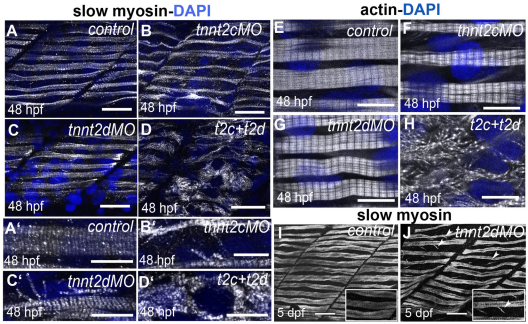
tnnt genes are required for sarcomere assembly in slow-twitch muscle. (A–D) Slow myosin staining (white) in 48 hpf control embryos (A,A′) and in embryos injected with tnnt2c MO (B,B′), tnnt2d MO (C,C′) or co-injected with tnnt2d and tnnt2c MOs (D,D′) shows that myofibrillar organisation is lost in the double-injected embryos. Nuclei are counterstained with DAPI (blue). A–D are lateral views of rostral somites and A′–D′ are higher magnification images showing muscle cells. In both cases, confocal images are from the most superficial layer of trunk muscles. Scale bars: 25 μm (A–D); 10 μm (A′–D′). (E–H) Phalloidin staining (white) in 48 hpf control embryos (E) and in embryos injected with tnnt2c MO (F), tnnt2d MO (G) or co-injected with tnnt2d and tnnt2c MOs (H) confirms loss of sarcomeres in double-injected embryos. Nuclei are counterstained with DAPI (blue). Scale bars: 10 μm. (I–J) Slow myosin staining (white) of control (I) and tnnt2d MO-injected (J) embryos shows fairly normal slow fibres at 5 dpf in the MO-injected embryo, with some myofibrils detached from the main bundles (white arrowheads). Scale bars: 30 μm, insets are 2× magnifications of corresponding panels.

