Fig. 6.
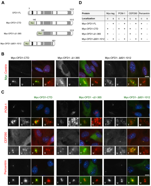
Ciliopathy protein localizations upon expression of truncated OFD1 constructs. (A) Schematic representation of Myc-tagged OFD1 deletion constructs. Coiled-coil (grey) and the LisH (black) motif, as well as amino acids numbers, are indicated. (B) hTERT-RPE1 cells were transfected with Myc-tagged OFD1 constructs as indicated and co-stained with antibodies against the Myc tag (greyscale and green in merge) to determine the localization of recombinant OFD1 protein and centrin2 (red in merge). Enlargements show centrosomal regions in transfected (T) and untransfected (U) cells. (C) hTERT-RPE1 cells were transfected with Myc-tagged OFD1 constructs as indicated and co-stained with antibodies against the Myc tag (green) and against PCM-1, CEP290 or pericentrin (greyscale and red in merge). Enlargements show centrosomal regions in transfected and untransfected cells. Merge images including DNA (blue) are shown in B and C. Scale bars: 10 μm. (D) The primary localization (+ present; − absent) of the Myc–OFD1 protein, PCM-1, CEP290 and pericentrin in the presence of the different OFD1 constructs is indicated. Results for each stain are based on at least 100 transfected cells for Myc-OFD1 full-length (FL), C-terminal domain (CTD) and Δ1-365, and 25 cells for Myc-OFD1 Δ601-1012, which transfected with much lower efficiency. c, centrosomes; s, centriolar satellites.

