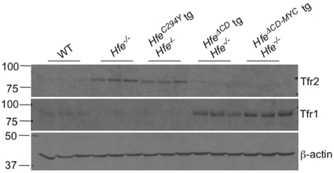
Figure 6.
Tfr1 and Tfr2 protein expression in Hfe−/−Hfe-transgenic animals. Liver protein lysates were analyzed for Tfr2 (top panel) or Tfr1 protein (middle panel) in 8-week-old wild-type (WT), Hfe−/−, Hfe−/− HfeC294Y, Hfe−/− Hfe-truncated transgenic (HfeΔCD tg), or Hfe−/− Hfe-cMyc transgenic (HfeΔCD−MYC tg) animals by Western blot. Equivalent loading of liver lysates was confirmed by immunoblot analysis using anti–β-actin antibody (bottom panel).

