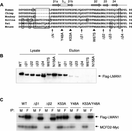
Figure 2.
The first β-sheet of the CRD is the binding motif for MCFD2. (A) Alignment of the first 56 amino acids of the mature human LMAN1 with the orthologs from chimp, monkey, bovine, rat, and mouse. Amino acid residues that differ from the consensus are boxed. The secondary structures are indicated on top of the sequences. The locations of deletions (arrows) and point mutations (arrow heads) used in the study are denoted under the sequences. (B) Mannose binding activities of different LMAN1 mutants. COS1 cells were transfected with the wild-type and the indicated LMAN1 mutants. LMAN1 proteins that are in the cell lysate and that are eluted from the mannose agarose beads are detected by Western blot analysis. Lysate lanes represent 20% of the input added to the mannose beads. (C) Co-IP of LMAN1 mutants with MCFD2. COS1 cells were cotransfected with Flag-tagged LMAN1 mutants and myc-tagged wild-type MCFD2. Cell lysates were immunoprecipitated with anti-myc for MCFD2 and anti-Flag for LMAN1.

