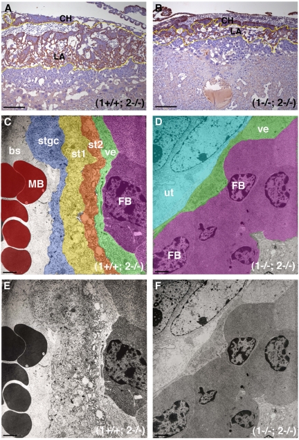
Figure 4.
MT1-MMP/MT2-MMP deficiency leads to disruption of LA architecture. (A) Control placenta from E10.5 embryo stained for HAI-1. Note the localization of brown immunoreactivity in CHs and in the differentiated trophoblasts of the LA outlined in yellow. (B) The staining pattern in a (1−/−; 2−/−) littermate is confined to a more restricted area due to the poor development of the LA (yellow outline). (C) Ultrastructure of the LA from a control placenta demonstrating the trilaminar structure of the fetal vasculature. Pseudocolors show the fetal-maternal interface composed of the fetal vascular endothelium (ve, green), 2 layers of syncytial trophoblasts (st2, orange and st1, yellow) and the STGC lining the MSs (stgc, blue). Fetal blood cell (FB, purple), maternal blood cell (MB, red). Note fetal red cells are nucleated in contrast to maternal cells. (D) Pseudocolored electron microscope image shows that the 2 syncytial layers are missing in the double-deficient placenta and the MSs are not in proximity. The fetal vascular endothelium in green (ve) is surrounded by undifferentiated trophoblasts in cyan (ut). Fetal blood cells (FB, purple). (E) Unaltered version of image shown in panel C. (F) Unaltered version of image shown in panel D. Scale bars: (A-B) 200 μm; (C-F) 2 μm.

