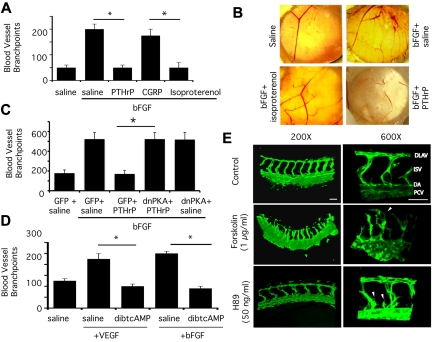
Figure 1.
PKA activation suppresses angiogenesis. (A) Mean vessel branchpoints ± SEM in CAMs of 10-day-old chicken embryos stimulated with saline or bFGF and topically treated with saline, 20μM PTHrP, calcitonin gene related peptide (CGRP), or isoproterenol, n = 10, *P < .05. (B) Images of CAMs stimulated with saline or bFGF and treated with saline, PTHrP, or isoproterenol. (C) Mean vessel branchpoints ± SEM in chorioallantoic membranes of 10-day–old chicken embryos stimulated with saline or bFGF, transfected with N1-GFP or pcDNA 3.1 V5/his dnPKA (dominant negative PKA = mutationally inactive PKA regulatory subunit I) as previously described10 and topically treated with saline or PTHrP, n = 10, *P < .05. (D) Mean vessel branchpoints ± SEM in chorioallantoic membranes of 10-day-old chicken embryos stimulated with saline, VEGF-A or bFGF, and topically treated with dibutyryl cAMP or saline, n = 10, *P < .05. (E) Images of 32 hpf transgenic GFP-expressing zebrafish larvae in vasculature [TG(fli1:EGFP1)] after treatment for 24 hours with forskolin or H89, a PKA inhibitor. DA (dorsal aorta), PCV (posterior cardinal vein), ISVs (intersegmental vessels), and DLAVs (dorsal longitudinal anastomotic vessels) are indicated for control-treated embryos.

