Figure 6.
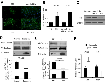
PKA and Src oppositely regulate VE-cadherin function. (A) Anti–VE-cadherin immunostaining of endothelial cell monolayers cultured in the presence of the Src inhibitor PP2 (PP2) or chemically inert control PP3 (PP3) and of endothelial cells transfected with Src siRNA or control siRNA. (B) Quantification of the mean ± SEM percent adherent cells in A. (C) Immunoblotting to detect Src of endothelial cells expressing Src siRNA or scrambled siRNA. (D) Immunoblot of phosphoVE-cadherin, VE-cadherin, and β-catenin after treatment of VEGF-stimulated endothelial cells with culture medium or forskolin. Graphs: ratios of phosphoVE-cadherin or β-catenin to total VE-cadherin. (E) Immunoblot of phosphoVE-cadherin, VE-cadherin, and β-catenin after treatment of VEGF-stimulated endothelial cells with PP2 or PP3. Graphs: ratios of phosphoVE-cadherin or β-catenin to total VE-cadherin. (F) Cell-cell adhesion of GFP or SrcA transfected EC treated with or without forskolin, expressed as mean ± SEM percent adherent cells.

