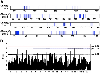
Figure 4.
Interval-specific haplotype analysis and HAM. (A) Each QTL was reduced by interval-specific haplotype analysis of the strains involved to the intervals indicated by the blue bars, prioritizing regions most likely to contain the QTL gene. QTL names and Chr locations are given to the left, with Chr positions (Mb) below. (B) Example of results obtained by HAM. Positions of the most significant peaks within the common combined cross/interval-specific haplotyping interval are given in Table 4.

